31 Jan What is the Hippocampus and Why Do I Hear About it in SUDC Research?
To say our central nervous system (CNS) is complex is an understatement. It comprises both the brain and the spinal cord with billions of cells, called neurons, forming connections all throughout that help us transmit information from the top-down (brain to the body) and from the bottom-up (body to the brain). Though complex, the brain can be organized in different parts that work synchronously to perform specific functions. There is the cerebrum, which is the largest part of the brain that controls all the higher functions: synthesizing our five senses, speech, comprehension, reasoning, emotions, learning, and movement control. The cerebellum, located right below the cerebrum, is responsible for coordinating muscle movements, retain your posture, and keep your balance. Finally, the brainstem is how the cerebrum and cerebellum relay information to the spinal cord. It is critical in many autonomic functions, or actions that you do not need to consciously think about to perform, such as maintaining breathing, heart rate, body temperature, sleep, digestion, and much more.
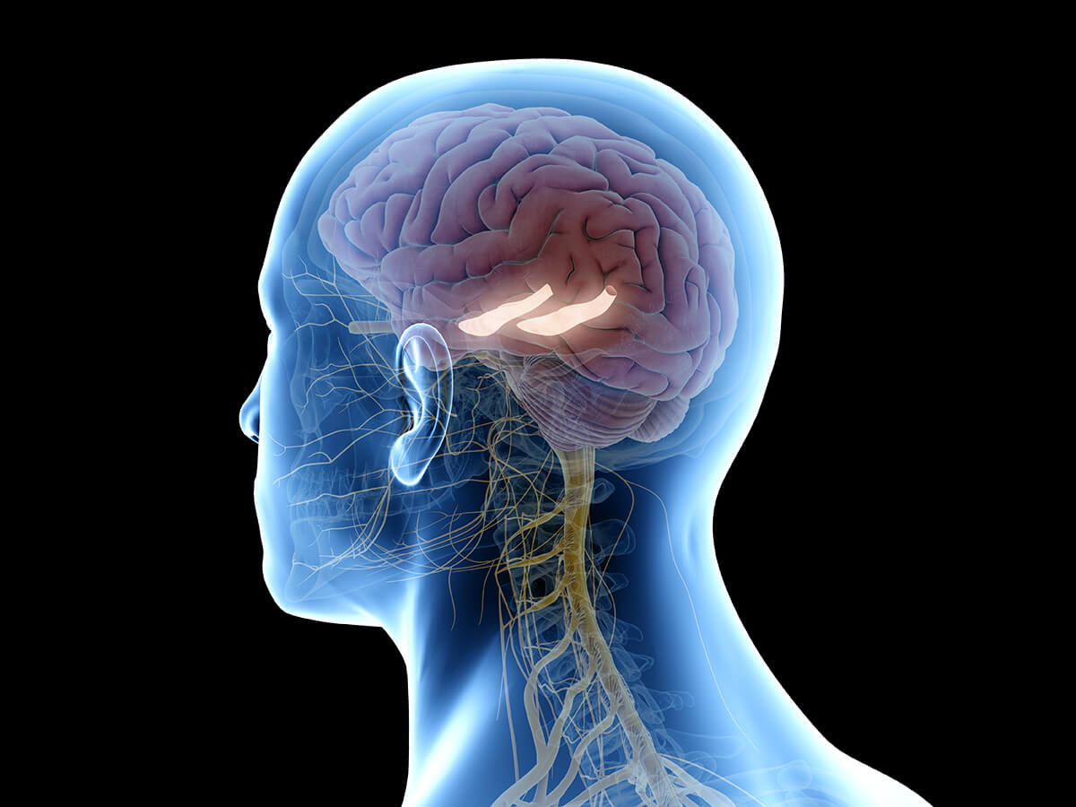
The cerebrum, with both the right and left hemispheres, can be further divided into the cortex and the deep structures. The cortex is separated into different lobes: frontal, temporal, parietal, and occipital. Each of these lobes is responsible for specific functions, but they also communicate with each other for more complicated functions. Deep to the cerebrum are numerous structures, containing more sub-structures, that further contribute to several bodily functions. These structures are the hypothalamus, pituitary gland, pineal gland, thalamus, basal ganglia, and limbic system. The limbic system, the part of the brain largely responsible for our emotions and behaviors, includes the structure of interest in this article: the hippocampi (singular: hippocampus).
The hippocampus is named after the Latin words hippos (horse) and kampos (sea monster) because its shape resembles the tail of a sea horse. Our brain has two hippocampi, with one in each hemisphere of the brain. They play a key role in creating and storing both our short-term and long-term memories. The hippocampus, when dissected and examined on glass histology slides, is further organized into distinct parts comprising separate groups of neurons that communicate with each other and with neurons in other parts of the brain. When the hippocampi are damaged, memory formation and retrieval can both be affected.
In addition to their role in memory, the hippocampi are involved in mesial temporal lobe epilepsy (MTLE), a specific type of temporal lobe epilepsy (most common form of epilepsy comprising around 60% of all epilepsy cases).1 MTLE is a focal epilepsy because its seizures can be traced back to specific brain structures medial to, or deep to, the temporal lobe, which includes the hippocampi and surrounding areas. Therefore, damage to this area of the brain, such as in the case of brain injury, tumors, or congenital malformations may increase the risk of MTLE in people. Another risk factor of MTLE is a history of febrile seizures; however, it is important to note that the vast majority of people with febrile seizures do not develop temporal lobe epilepsy.1 The risk of developing epilepsy of any kind following a febrile seizure is 2.0-7.5% whereas the risk following a complex febrile seizure increases to 10-20%.2
Since febrile seizures have also been associated with some SUDC research, the hippocampi became an area of interest in both SIDS and SUDC research, with some studies identifying hippocampal findings in some cases of sudden unexplained deaths in infants and children.3-5 In June 2021, Leitner et al. published the first ever blinded review of hippocampal slides from both SUDC and SEDC (sudden explained death in childhood) cases. In the comprehensive review, nine board-certified pathologists examined microscopic hippocampal sections without knowledge of which cases were SUDC or which were SEDC. The reviewers found that about half of both groups had findings considered ‘abnormal’. They had little agreement and high variability between the reviewers. By highlighting the lack of consistent evidence that linked hippocampal abnormalities to SUDC, the authors emphasized discrepancies in previously published studies. As with other areas of SUDC research, more research is warranted to establish standards in both evaluating hippocampal histology and recognizing the normal variations and changes that are related to SUDC or febrile seizures.6
Written by:
Joyce H. Lee, MS
Medical College of Wisconsin, Class of 2024
Former Research Data Associate at the SUDC Registry and Research Collaborative (2017-2020)